Medial Epicondyle Of Femur Muscle Attachments / Bones Of The Lower Limb Anatomical Basis Of Injury : Head, medial larger epicondyle, lateral olecrenon fossa, distal.. The common extensor tendon attaches to the lateral epicondyle, acting as the common attachment for the superficial extensor muscles of the forearm. The condition is caused by the irritation or compression of the proximal sciatic nerve by the piriformis muscle, which connects the sciatic notch with the greater trochanter of the femur. All superficial muscles are arises from the medial epicondyle of humerus but they are inserted into the different part except. Head lateral epicondyle medial epicondyle olecrenon fossa. The medial condyle is one of the two projections on the lower extremity of femur, the other being the lateral condyle.
• the joined tendons of the sartorius, gracilis, semitendinosus muscles cross on top of the lower part of the. Located above the medial condyle, it bears an elevation, the adductor tubercle, which serves for the attachment of the superficial part, or tendinous insertion. A medial epicondyle fracture is an avulsion injury of the attachment of the common flexors of the forearm. Important features of this bone include the head, medial and lateral condyles, patellar surface, medial and lateral epicondyles, and greater and lesser the epicondyles and trochanters are all important attachment sites for various muscles. Lateral epicondyle of the femur.
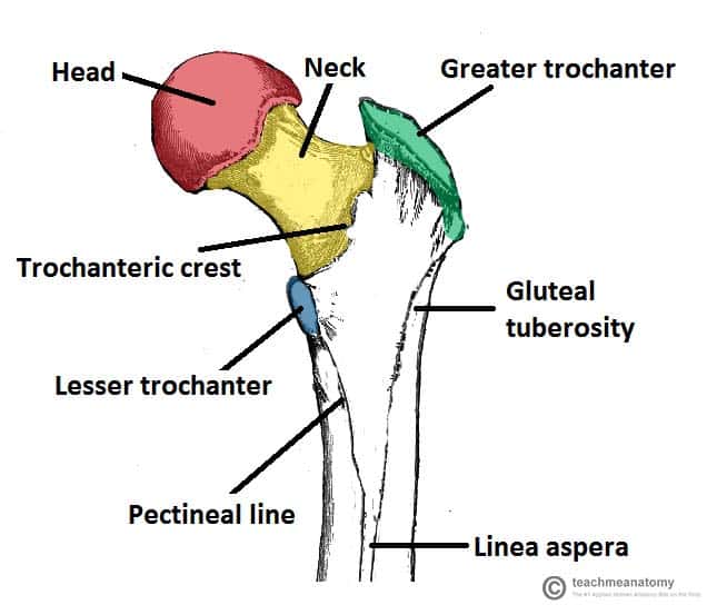
The Femur Proximal Distal Shaft Teachmeanatomy from teachmeanatomy.info All 4 muscles have a common origin at the medial epicondyle of the humerus, known as the common flexor tendon. It acts as the site of origin and attachment of many muscles and ligaments, and can be divided into three parts; A medial epicondyle fracture is an avulsion injury of the attachment of the common flexors of the forearm. Located above the medial condyle , it bears an elevation, the adductor tubercle , 1 which serves for the attachment of the superficial part, or tendinous insertion, of the adductor magnus. Medial enlargement of distal end; Medial condyle is convex medially. Olecrenon process, proximal radial notch, lateral styloid process, towards 'pinky'. What's actually happening is a degeneration of the tendons that connect to your medial epicondyle.
Abduction and medial rotation of femur. The femur is the only bone in the thigh and the longest bone in the body. All superficial muscles are arises from the medial epicondyle of humerus but they are inserted into the different part except. From wikipedia, the free encyclopedia. It also bears a prominent point called the medial epicondyle. Tennis elbow management a multimodal management. Medial and lateral intermuscular septa are attached to the lips of the linea aspera and to the supracondylar line. The medial epicondyle of the femur is an epicondyle, a bony protrusion, located on the medial side of the femur at its distal end. This condition is characterized by irritation and inflammation of the growth plate (apophysis) on the inner side of the elbow (medial epicondyle). At its upper part is the adductor tubercle and behind it is a. All 4 muscles have a common origin at the medial epicondyle of the humerus, known as the common flexor tendon. The medial and lateral epicondyles are small bony tuberosities on the distal end of the humerus (fig. It also provides attachment for the tendon of adductor magnus.
Tennis elbow management online course: The femur is the only bone located within the human thigh. The condition is caused by the irritation or compression of the proximal sciatic nerve by the piriformis muscle, which connects the sciatic notch with the greater trochanter of the femur. This condition is characterized by irritation and inflammation of the growth plate (apophysis) on the inner side of the elbow (medial epicondyle). Repetitive overuse of the tendons.
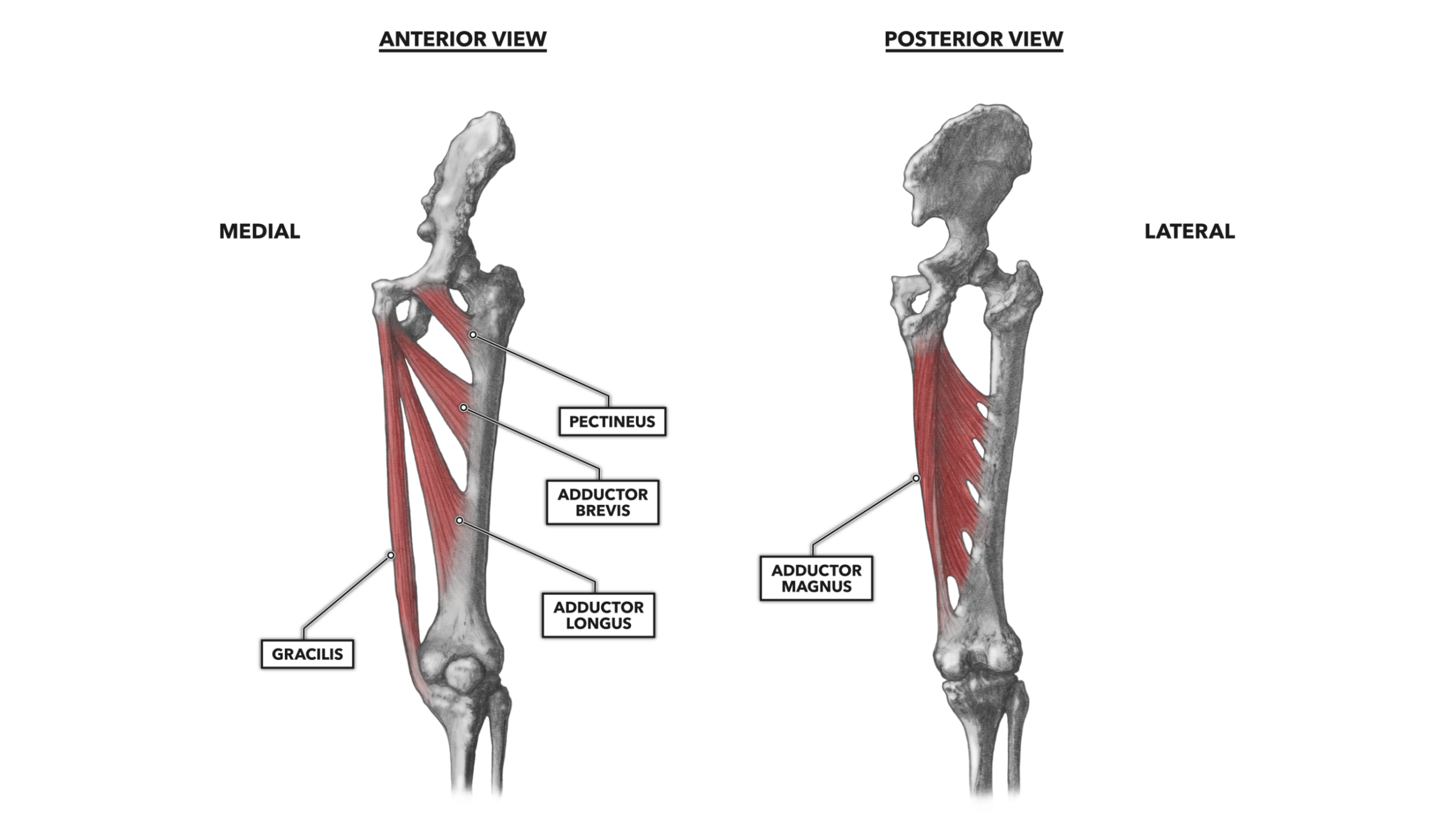
Crossfit Hip Musculature Part 4 Medial Muscles from www.crossfit.com Does not contribute to longitudinal growth (apophysis). Repetitive overuse of the tendons. For medial epicondyle fractures, nonoperative management of fractures displaced up to 15 mm does not the medial epicondyle is a prominent palpable process that projects medially from the trochlea and is the attachment of medial collateral ligament components is pictured. Head, medial larger epicondyle, lateral olecrenon fossa, distal. Fasical attachments at pubic symphysis. Medial and lateral intermuscular septa are attached to the lips of the linea aspera and to the supracondylar line. The medial collateral ligament extend from the medial epicondyle of the femur to below the medial condyle of the tibia. That force and overuse has started to add up and the area is no longer just inflamed, but degenerated.
Femoral head with the acetabulum of the pelvis knee:
All 4 muscles have a common origin at the medial epicondyle of the humerus, known as the common flexor tendon. Smooth surface articulates with tibia and patella comment: The common extensor tendon attaches to the lateral epicondyle, acting as the common attachment for the superficial extensor muscles of the forearm. Related online courses on physioplus. Posterior aspect of medial femoral condyle. The medial collateral ligament extend from the medial epicondyle of the femur to below the medial condyle of the tibia. Some of the most discussed epicondyles are the medial and lateral epicondyles of femur and humerus. Important features of this bone include the head, medial and lateral condyles, patellar surface, medial and lateral epicondyles, and greater and lesser the epicondyles and trochanters are all important attachment sites for various muscles. A medial epicondyle fracture is an avulsion injury of the attachment of the common flexors of the forearm. Providing a surface for attachment of muscle and ligament is the main function of the epicondyle. Displacement is difficult to measure accurately as medial epicondyle is located on the posteromedial aspect of the distal humerus and fragment displaces. All superficial muscles are arises from the medial epicondyle of humerus but they are inserted into the different part except. The condition is caused by the irritation or compression of the proximal sciatic nerve by the piriformis muscle, which connects the sciatic notch with the greater trochanter of the femur.
Abduction and medial rotation of femur. From the femoral region, they extend obliquely across the medial side of the knee's anterior, between the medial this motion is assisted by the contraction of the other muscles of the quadriceps femoris group, which simultaneously pull on the tibia to extend the knee. The femur is the longest and strongest bone of the body, present in the thigh (latin femur = thigh). Femoral head with the acetabulum of the pelvis knee: Posterior aspect of medial femoral condyle.
Leg Knee Anatomy from assets.website-files.com Fasical attachments at pubic symphysis. Provides attachment for medial head of gastrocnemius and adductor magnus muscles, and tibial collateral ligament of knee; The medial and lateral epicondyles are small bony tuberosities on the distal end of the humerus (fig. Located above the medial condyle, it bears an elevation, the adductor tubercle, which serves for the attachment of the superficial part, or tendinous insertion. That force and overuse has started to add up and the area is no longer just inflamed, but degenerated. Medial intermuscular septum is connected to the medial lip of its most notable point is named medial epicondyle, which gives connection to the upper end of. These septae separate the extensor muscles from the adductor medially, and. It acts as the site of origin and attachment of many muscles and ligaments, and can be divided into three parts;
The condition is caused by the irritation or compression of the proximal sciatic nerve by the piriformis muscle, which connects the sciatic notch with the greater trochanter of the femur.
Some of the most discussed epicondyles are the medial and lateral epicondyles of femur and humerus. In primitive tetrapods, the main points of muscle attachment along the femur are the internal trochanter and third trochanter, and a ridge along the ventral surface of the femoral shaft. Lateral and medial condyles of the femur with the tibial plateaus of the tibia (tibiofemoral joint) the medial epicondyle is situated below and anterior to the adductor tubercle. The medial epicondyle is the attachment site for the forearm muscles used in throwing and helps to stabilize the elbow during the throwing motion. Additionally, the ventral and dorsal epicondyles of birds are important for them to keep. Does not contribute to longitudinal growth (apophysis). The medial epicondyle of the femur is an epicondyle, a bony protrusion, located on the medial side of the femur at its distal end. The common extensor tendon attaches to the lateral epicondyle, acting as the common attachment for the superficial extensor muscles of the forearm. Abduction and medial rotation of femur. It also bears a prominent point called the medial epicondyle. Posterior aspect of medial femoral condyle. A medial epicondyle fracture is an avulsion injury of the attachment of the common flexors of the forearm. Olecrenon process, proximal radial notch, lateral styloid process, towards 'pinky'.
Medial intermuscular septum is connected to the medial lip of its most notable point is named medial epicondyle, which gives connection to the upper end of medial epicondyle femur. Tennis elbow management online course:

0 Comments:
Posting Komentar